Liver Ultrasound: A Detailed Overview
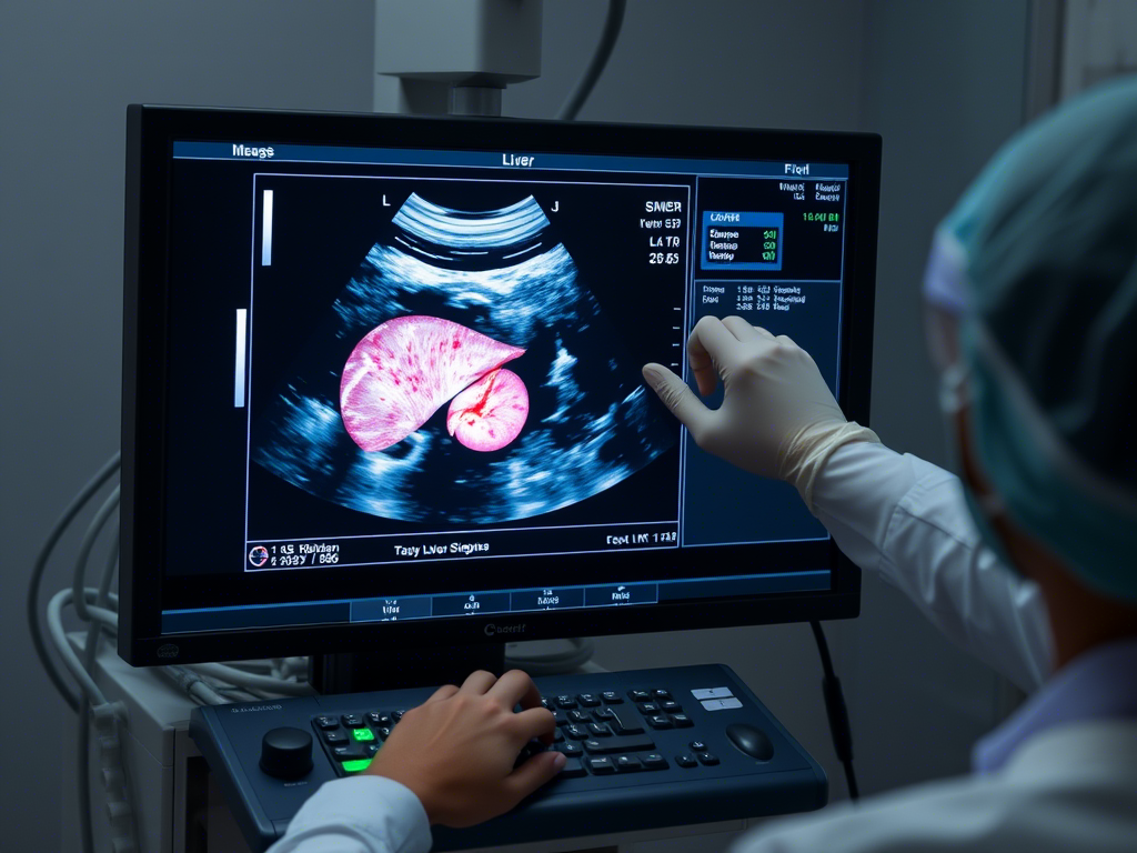
A Liver Ultrasound is a non-invasive imaging test that uses high-frequency sound waves to create real-time images of the liver and surrounding structures. It helps assess liver size, shape, texture, and detect abnormalities such as fatty liver, cysts, tumors, cirrhosis, and liver infections.
How a Liver Ultrasound Works
The procedure involves:
- Applying a gel to the abdomen to improve sound wave transmission.
- Moving a handheld transducer over the upper right abdomen.
- Capturing images as sound waves bounce off liver tissues.
- The test is painless, radiation-free, and takes about 15-30 minutes.
Uses of a Liver Ultrasound
1. Detecting Fatty Liver Disease
- Identifies fat accumulation in the liver, which may lead to complications if untreated.
2. Diagnosing Liver Cirrhosis
- Helps detect scarring and damage caused by chronic liver disease.
3. Identifying Liver Tumors or Cysts
- Differentiates between benign and malignant growths.
4. Evaluating Hepatitis and Liver Infections
- Assesses liver swelling and inflammation due to hepatitis or other infections.
5. Checking for Bile Duct Obstruction
- Detects blockages, gallstones, or bile duct narrowing affecting liver function.
Benefits of a Liver Ultrasound
- Non-invasive and painless.
- Radiation-free and safe for all ages.
- Provides real-time imaging for accurate diagnosis.
Conclusion
A Liver Ultrasound is a crucial tool for diagnosing fatty liver, cirrhosis, tumors, and infections, ensuring early detection and effective treatment.


Reviews
There are no reviews yet.