MRI Lumbar Spine: A Detailed Overview
An MRI Lumbar Spine is a non-invasive imaging test that provides detailed visuals of the bones, discs, nerves, muscles, and soft tissues in the lower back (lumbar region). It helps diagnose conditions such as herniated discs, spinal stenosis, nerve compression, arthritis, and spinal tumors, aiding in effective treatment planning.
How an MRI Lumbar Spine Works
MRI (Magnetic Resonance Imaging) uses a strong magnetic field and radio waves to generate high-resolution images of internal structures. Unlike X-rays or CT scans, MRI does not use radiation, making it a safe and effective imaging technique, especially for soft tissue evaluation.
Procedure:
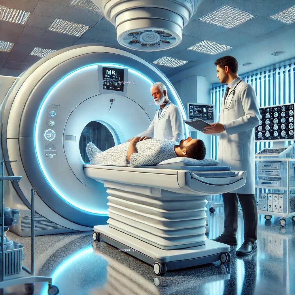
- The patient lies on a movable table, which slides into the MRI scanner.
- The scan typically takes 30 to 45 minutes, during which the patient must remain still for clear imaging.
- In some cases, a contrast dye (gadolinium) may be injected to enhance visualization of certain structures.
- The procedure is painless and requires no recovery time.
Uses of an MRI Lumbar Spine Scan
1. Diagnosing Herniated or Bulging Discs
- Detects disc herniation or bulging, which can press on nerves and cause back pain, numbness, or weakness in the legs.
- Helps evaluate degenerative disc disease, which occurs due to aging or injury.
2. Identifying Spinal Stenosis and Nerve Compression
- Assesses spinal canal narrowing (stenosis), which can compress nerves and lead to pain, tingling, or difficulty walking.
- Helps detect sciatica, a condition where the sciatic nerve is compressed, causing pain radiating down the legs.
3. Detecting Spinal Cord and Nerve Abnormalities
- Identifies nerve inflammation, spinal cord injuries, or infections that may cause back pain or neurological symptoms.
- Helps diagnose multiple sclerosis (MS) or other spinal cord diseases.
4. Evaluating Arthritis and Degenerative Changes
- Detects osteoarthritis, rheumatoid arthritis, and spondylosis, which cause stiffness, pain, and reduced mobility.
- Helps assess facet joint degeneration, which can contribute to chronic lower back pain.
5. Identifying Spinal Tumors and Infections
- Detects benign or malignant tumors, infections (osteomyelitis), or abscesses in the lumbar spine.
- Helps evaluate unexplained lower back pain that doesn’t improve with standard treatments.
Benefits of an MRI Lumbar Spine
- High-resolution imaging for accurate diagnosis.
- No radiation exposure, making it a safer alternative to CT scans.
- Early detection of spinal conditions, preventing long-term complications.
- Essential for pre-surgical evaluation and post-treatment monitoring.
Safety and Considerations
- Generally safe and painless.
- Not suitable for individuals with metal implants, pacemakers, or severe claustrophobia.
- Contrast dye (if used) is typically safe but should be avoided in patients with severe kidney disease or allergies.
Conclusion
An MRI Lumbar Spine is a crucial diagnostic tool for evaluating lower back pain, nerve compression, herniated discs, arthritis, and spinal abnormalities. It provides detailed, radiation-free imaging, ensuring early diagnosis and effective treatment planning for better spinal health and mobility.

Reviews
There are no reviews yet.